


Animal Multi-modal Microcatheter Endoscopy GPA-US-10, GUS-2D/3D, GOCT-OCT, GOCT-US-10 lMulti-modal (Photoacoustic, Ultrasonic, OCT) in vivo imaging lPhotoacoustic lateral direction Resolution down to 15μm, OCT lateral direction Resolution down to 8μm, Ultrasound imaging depth up to 30mm 3D Merged Imaging
Product Advantages
Defects of Traditional Optical Endoscopes
The in - vivo endoscopic imaging depth is shallow and it is unable to visualize structural lesions.
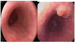
White-light endoscopy
It has high resolution but shallow penetration;Mucosal lesions
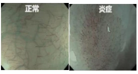
Narrow - band endoscopy
It has high resolution and shallow penetration;Mucosal lesions
Innovative multimodal endoscope
In vivo imaging; larger depth; detect mucosal lesions and structural lesions.
Multimodal endoscopy
Higher resolution and deeper penetration
Penetration:
The whole biological tube wall
Resolution:
Photoacoustic transverse≤15μm, axial≤65μm
Ultrasound: axial:≤65μm
OCT: transverse≤20μm,axial≤8μm
Highlights:
OCT Imaging
Ultrasound imaging
Photoacoustic imaging
Structural imaging
Functional imaging
High resolution, deep penetration, three-dimensional imaging, specific imaging
Structural lesions, mucosal lesions


Penetration: The full thickness of the biological tube wall.
Resolution: Increased by 20 times.


Applications
Meet the endoscopic needs of various experimental animals
Endoscopic experiments on rats,mice,rabbits and pigs


Product Description
The photoacoustic multi-modal imaging apparatus integrates optical imaging and acoustic imaging methodologies to generate high-resolution pictures of biological tissues at diverse depths. This technique can be utilized in multiple sectors, including cancer diagnosis, brain imaging, and vascular imaging. The photoacoustic multi-modal imaging system possesses benefits like being non-invasive, enabling real-time imaging, and having a low cost, which renders it a prospective instrument for medical research and clinical uses.
Product Parameters
There are four models of the Animal Multimodal Microcatheter Endoscope Imaging System. Please check the parameters as follows.
|
Product Model |
GPA-US |
GUS |
GOCT-US |
G0CT |
|
Imaging modality |
PAE&US |
US |
OCT&US |
OCT |
|
Application |
Cardiovascular, digestive system, otolaryngology |
Digestive system, ear, nose and throat, urinary system |
Cardiovascular, Digestive System, ENT |
Cardiovascular, Digestive System, Ear, Nose and Throat, Cranial Nervous System |
|
Scan angle/ Retraction distance |
360°/80 mm |
360°/40 mm |
360°/ 80 mm |
360°/80 mm |
|
Scan mode |
2D/3D |
2D/3D |
2D/3D |
2D/3D |
|
Probe frequency |
12/20 MHz |
12/20 MHz |
12/20 MHz |
/ |
|
Imaging speed |
30 fps |
30 fps |
30 fps |
100 fps |
|
Resolution |
(PAE) transverse≤15μm,axial≤65μm (US)axial≤65μm |
axial:≤65μm |
(OCT) transverse≤20um,axial≤8um (US)axial≤65μm |
transverse≤20um, axial≤8 um |
|
Imaging depth |
PAE≥3mm US≥30 mm |
≥30 mm |
OCT≥ 3mm US≥30 mm |
≥3mm |
|
Catheter diameter |
1.0/2.5 mm |
0.7/2.1/2.5 mm |
1.0/2.5 mm |
1.0 mm |
|
Tube selection |
Standard, balloon, guidewire |
Standard, balloon, guidewire |
Standard, balloon, guidewire |
Standard, balloon, guidewire |
Customer Pain Points & Solutions - Imaging Examples

Customer Pain Point : It is difficult to perform endoscopic imaging of the intravascular hierarchical structure and plaques.
East Solution: Non-invasive, label-free, and ultra-high-resolution visualization of intravascular plaques and hierarchical structures.
Through the consistency evaluation of multimodal endoscopic imaging and histology, the diagnostic ability of intravascular photoacoustic imaging for lipids has been verified.
IVPA/US Images and Stained Images of the Rabbit Abdominal Aorta


Consistency Evaluation of Photoacoustic Imaging and Histology
Lei P et al., J Biophotonics. 2020 Dec;13(12):e202000162. DOI:10.1002/jbio.202000162.
Customer Pain Point: It is difficult to perform endoscopic imaging of the hierarchical structure within the digestive tract.
East Solution: Non-invasive, label-free, and ultra-high-resolution visualization of the hierarchical structure within the digestive tract.
Non-invasive, label-free, and ultra-high-resolution visualization of the vascular networks and hierarchical structures at different depths in the colorectum.
Photoacoustic (PA) Imaging of the Rabbit Colon and Rectum (Muscular Layer and Serosa, Submucosa, Mucosa)

Xiong K et al., Optics Letters, 2019, 44(11):2681.DOI:10.1364/ol.44.002681.
Imaging Examples

Scanning and imaging of the large lumens (such as the esophagus, trachea, intestine, stomach wall, etc.) with a balloon catheter endoscope.
Ultrasound imaging of the colorectum in living pigs.


Scanning and imaging with a standard catheter endoscope.
Optical Coherence Tomography (OCT) imaging of the coronary arteries of the porcine heart.
3D Optical Coherence Tomography (OCT) images of the coronary arteries of the porcine heart.


Our Factory
Shenzhen East Scientific Instrument Co., Ltd. is a highly innovative high - tech enterprise. It is committed to the innovative development of global microbiology research and application and the application of optical imaging technology in the field of life sciences. The company is led by academicians of the American Academy of Microbiology, in conjunction with the Graduate School at Shenzhen, Tsinghua University, Southern University of Science and Technology, South China Normal University, etc. It has brought together many multidisciplinary experts in microbiology, mechanical automation, electronic informatics, etc. from both domestic and foreign countries, and has a group of high - quality young technical backbones. It has a complete optical detection experimental platform and cross - integrates the laboratory equipment industry and the Internet industry.
Hot Tags: animal multimodal microcatheter endoscope imaging system, China animal multimodal microcatheter endoscope imaging system manufacturers, suppliers, factory, multi modal imaging for educational applications, multi modal imaging for benchmarking, multi modal imaging for community engagement, multi modal imaging for automotive research, multi modal imaging for compliance, Multimodal Endoscopic Imaging System


